 Cancer as a Metabolic Disease
 by Jeffrey Dach MD
Losing the War On Cancer
 We
have lost the war against cancer. This becomes obvious by taking a
quick look at the chart below. Cancer Death Rates have declined
slightly since 1975 due to reduction in lung cancer rates from decreased
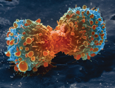
cigarette consumption. Otherwise, there has been little change. Above left lung cancer cell dividing courtesy of NIH .
We
have lost the war against cancer. This becomes obvious by taking a
quick look at the chart below. Cancer Death Rates have declined
slightly since 1975 due to reduction in lung cancer rates from decreased
cigarette consumption. Otherwise, there has been little change. Above left lung cancer cell dividing courtesy of NIH . Below chart image courtesy of Scientific American.
The Failure of Chemotherapy
In 1997, John C. Bailar M.D. explained how we lost the "War Against Cancer" : "The failure of chemotherapy to control cancer has become apparent even to the oncology establishment."
Here is a quote from Dr. Haines in Lancet: "The misplaced battlefield analogy has led to 40 years of toxic and overly aggressive chemotherapy in incurable solid cancers for which no studies have shown that maximum tolerated doses of chemotherapy achieve longer survival or better quality of life than do minimum effective doses. This approach has led to inappropriate and toxic therapies for many patients.... "
Dr Carlos Garcia says cancer treatment with chemotherapy has a 97% failure rate: Three Per Cent Efficacy Of CHEMO On Cancer by Carlos Garcia MD.
Chemotherapy kills normal cells as well as cancer cells
 The
obvious flaw in cancer chemotherapy treatment is the non-specificity.
In other words, cancer chemotherapy kills cancer cells as well as normal
cells. This unwanted toxicity to normal cells accounts for the adverse
side effects of chemotherapy, namely nausea, vomiting, loss of
appetite, weight loss, bone marrow suppression with anemia, hair loss
etc.
The
obvious flaw in cancer chemotherapy treatment is the non-specificity.
In other words, cancer chemotherapy kills cancer cells as well as normal
cells. This unwanted toxicity to normal cells accounts for the adverse
side effects of chemotherapy, namely nausea, vomiting, loss of
appetite, weight loss, bone marrow suppression with anemia, hair loss
etc.Finding a Selective Cancer Treatment, Leaving Normal Cells Unharmed
 The optimal cancer treatment is one that kills cancer cells while leaving normal cells unharmed. Dr Seyfried's article, "Cancer as a metabolic disease " points the way to this goal.(1) Dr. Seyfried says: "cancer is primarily a metabolic disease involving disturbances in energy production through respiration and fermentation."(1)
Electron microscope studies have shown abnormalities in the
mitochondria of cancer cells.(2) The cancer cell's mitochondria have
undergone "metabolic reprogramming", thereby providing a key which can
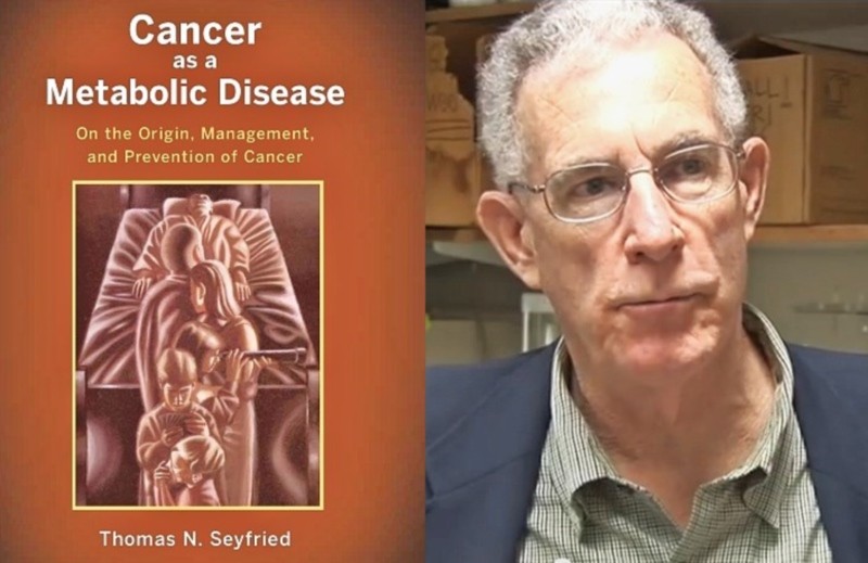
be exploited to devise a more selective cancer treatment. (3,4) Above left image, Cover of book Cancer as a Metabolic Disease by Dr Thomas Seyfried courtesy of the Examiner.
The optimal cancer treatment is one that kills cancer cells while leaving normal cells unharmed. Dr Seyfried's article, "Cancer as a metabolic disease " points the way to this goal.(1) Dr. Seyfried says: "cancer is primarily a metabolic disease involving disturbances in energy production through respiration and fermentation."(1)
Electron microscope studies have shown abnormalities in the
mitochondria of cancer cells.(2) The cancer cell's mitochondria have
undergone "metabolic reprogramming", thereby providing a key which can
be exploited to devise a more selective cancer treatment. (3,4) Above left image, Cover of book Cancer as a Metabolic Disease by Dr Thomas Seyfried courtesy of the Examiner. Drs Ko and Pederson, 3BP and Hexokinase II
Basic science studies by Drs Ko and Pedeson have uncovered the exact details of the metabolic reprogramming of the mitochondria of cancer cells. In cancer cells, an embryonic form of Hexokinase called Hexokinase II has been attached to the membrane pores of the mitochondria (called VDAC voltage dependent anion channels) . Hexokinase II is the first enzyme in glucose utilization and its location on the outer membrane pores allows for the massive utilization of glucose to feed the rapidly growing tumor mass. Separation of the Hexokinase II from the VDAC pore on the outer mitochondrial membrane triggers apoptosis (programmed cell death) of the cancer cell while sparing normal cells.(5,6)

Above image: Dr. Young Ko and Dr. Peter Pedersen with Dr. Humaira Khan, Medicor CEO (left) and Dr. Akbar Khan Medicor Medical Director (right) at the Medicor Office in Toronto, December 2008. Courtesy of Medicor.
Discovery of 3BP by Dr. Ko
Drs Ko and Pederson discovered a small molecule called 3BP which throws a "monkey wrench" into the metabolic machinery of the cancer cell, and induces apoptosis via separation of Hexokinase II from the outer mitochondrial membrane. (7)(27, 28)
3BP (3 Bromo Pyruvate) is a small non-toxic molecule that induces apoptosis in cancer cells while sparing normal cells, thus providing the most promising cancer treatment we have seen in many years. In-fighting over patent rights by the key players has delayed drug development and commercialization. Unfortunately, 3BP as a cancer treatment may never come to fruition. (7)
Natural Compounds that Disassociate Hexokinase II from VDAC
 Thankfully,
there are many other compounds in the natural world that act on this
same mechanism of selectively inducing apoptosis in cancer cells while
sparing normal cells. Many of these have already been commercialized
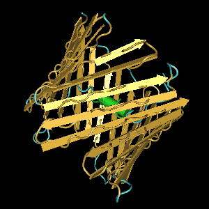
and are available at the health food store.Left Image VDAC courtesy of NIH.
Thankfully,
there are many other compounds in the natural world that act on this
same mechanism of selectively inducing apoptosis in cancer cells while
sparing normal cells. Many of these have already been commercialized
and are available at the health food store.Left Image VDAC courtesy of NIH.Resveratrol and Pterostilbenes
Resveratrol from grapes and its derivative Pterostilbene have been extensively studied and demonstrate striking anticancer activity. Studies show that Pterostilbene induces apoptosis via the mitochondrial pathway in breast cancer cell lines. See the article by Moon. Another article by Pei-Ching Hsiao. His study used acute myeloid leukemia cells, finding pterostilbene induced apoptosis in cancer cells vIa mitochondrial pathways, (with activation of caspase system). Another study by Alosi on Pterostilbene in Breast Cancer also showed similar findings with apoptosis induced by mitochondrial pathways. Another more recent study by Wang in 2012 showed Pterostilbene induces apoptosis and cell cycle arrest in breast cancer cells. See my previous article on this.
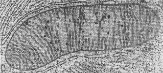
Abov image : electron microscope image of normal mitochondria courtesy of NIH.
Methyl Jasmonate
Methyl Jasmonate has been studied and found to disassociate Hexokinase II from the outer mitochondria membrane (at the VDAC) thus inducing apoptosis in cancer cells. Methyl jasmonate is ubiquitous in the plant world and found in the jasmine flower. It is used extensively by the fragrance industry for perfumes, and is available as one of many Jasmine teas.(8,9,10)
Oroxylin A - Chinese Skullcap
Oroxylin A is found in Chinese skullcap a medicinal plant, Scutellaria baicalensis Georgi. Researchers found Oroxylin A inhibits glycolysis and the binding of hexokinase II (HK II) with mitochondria in human breast carcinoma cell lines, thus inducing apoptosis (11,12)
Curcumin
Curcumin was studied by Dr Wang in an animal model of colorectal cancer showing Curcumin inhibits aerobic glycolysis and induces mitochondrial-mediated apoptosis through hexokinase II in human colorectal cancer cells in vitro. (13)(31) Curcumin is available at the health food store.
Betulinic acid
Betulinic acid naturally occurs in the bark of white birch trees and triggers mitochondrial apoptosis in cancer cells while sparing normal cells (14) Dr, Simone Fulda from Ulm Germany has written a number of important papers on targeting mitochondria in cancer cells with various drugs and natural compounds. (14,15)
See: Targeting_Mitochondria_Cancer_Therapy_Fulda_Nature_2010
Berberine - Oregon Grape
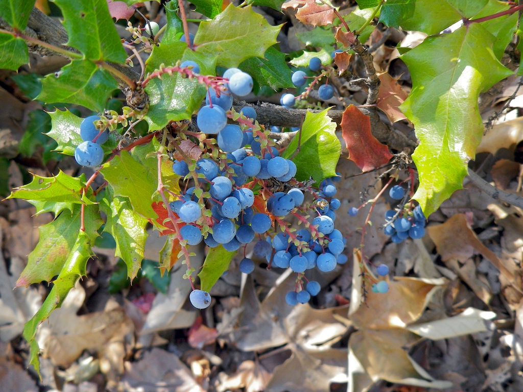 Berberine
derived from the Oregon Grape plant is widely available as a botanical
supplement at the health food store for blood sugar control. Berberine
also shows striking anti-cancer activity, inducing apoptosis via
mitochondrial pathways in numerous studies. Left Image Oregon Grape courtesy of wikimedia commons.(31,32) See this 2014 review in Molecules, Berberine as an "Epiphany Against Cancer". (32)
Berberine
derived from the Oregon Grape plant is widely available as a botanical
supplement at the health food store for blood sugar control. Berberine
also shows striking anti-cancer activity, inducing apoptosis via
mitochondrial pathways in numerous studies. Left Image Oregon Grape courtesy of wikimedia commons.(31,32) See this 2014 review in Molecules, Berberine as an "Epiphany Against Cancer". (32)Artemesinin (Chinese Wormwood)
An anti-malarial Chinese herb, artemesia, has been found to have profound anti-cancer activity against multiple cancer cell lines. Studies show induction of apoptosis through mitochondrial pathways. Approximately 400 studies have been published in the scientific literature in recent years.
Click on this link: Artemisinin_anti-Cancer_Publications, for the list of publications. Click on this link for an excellent review: Artemisinins_Krishna_Trends_Pharmacological_Science_2008
Vitamin K
Vitamin K has been available for many years with heath benefits in blood coagulation, bone density, and prevention of soft tissue calcification. See my previous article on Vitamin K. Another surprising benefit of Vitamin K is cancer prevention. A number of cell culture and animal xenograft studies shows that vitamin K2 induces apoptosis, programmed cell death, in cancer cells.(18-26) There were a number of different cancers studied including Glioblastoma, Hepatocellular Cancer, Lung Cancer, Prostate Cancer, etc.(18-26) Based on these reports, one might suggest adding Vitamin K to a supplement program for anyone seeking to prevent cancer, or cancer recurrence after treatment.
Buy Pterostilbene on Amazon
Buy Artemisinin
 on Amazon
Buy Berberine on Amazon
Buy Curcumin (Pure)
Buy Vitamin K (Synergy K)
Articles with related interest:
Nicholas Gonzalez and the Trophoblast Theory of Cancer
Iodine Treats Breast Cancer Overwhelming Evidence
Cannabis Oil Brain Tumor Remission
Natural Treatments for Skin Cancer
Addendum: Selectively killing cancer cells leaving normal cells unharmed - apoptosis through mitochondrial pathways
Mebendazole - Re purposing Old Drugs as Anti-Cancer Agents:
Researchers screened 2000 drugs currently approved for human use for anti-cancer activity against virulent melanoma cancer cell lines. They discovered mebenzadole as the most promising agent. "Mebendazole treatment induces apoptosis through the intrinsic and extrinsic (mitochondrial) pathways in melanoma cells but not in melanocytes... After treatment with 0.5 μmol/L mebendazole for 14 h, we observed overall microtubular network disarray in melan-a, M-14, and SK-Mel-19 cells, characterized by diffuse staining" .(33-37)
See: mebendazole metastatic colon cancer Nygren Acta Oncologica 2014
See Anti- Cancer Agents in Fruits and Vegetables:
Molecular targets of dietary agents prevention cancer aggarwal 2006
Jeffrey Dach MD
7450 Griffin Road Suite 190
Davie, Fl 33314
954-792-4663
link to this article:http://wp.me/p3gFbV-2p8
Links and References
1) http://www.ncbi.nlm.nih.gov/
Carcinogenesis. 2014 Mar;35(3):515-27.
Cancer as a metabolic disease: implications for novel therapeutics.
Seyfried TN1, Flores RE, Poff AM, D'Agostino DP.
1Biology Department, Boston College, Chestnut Hill, MA 02467, USA and.
Emerging evidence indicates that cancer is primarily a metabolic disease involving disturbances in energy production through respiration and fermentation. The genomic instability observed in tumor cells and all other recognized hallmarks of cancer are considered downstream epiphenomena of the initial disturbance of cellular energy metabolism. The disturbances in tumor cell energy metabolism can be linked to abnormalities in the structure and function of the mitochondria.
Cancer growth and progression can be managed following a whole body transition from fermentable metabolites, primarily glucose and glutamine, to respiratory metabolites, primarily ketone bodies.
2) http://www.ncbi.nlm.nih.gov/
Int J Biochem Cell Biol. 2009 Oct;41(10):2062-8.
Electron microscopy morphology of the mitochondrial network in human cancer.
Arismendi-Morillo G.
Mitochondria have been implicated in the process of carcinogenesis, which includes alterations of cellular metabolism and cell death pathways. The aim of this review is to describe and analyze the electron microscopy morphology of the mitochondrial network in human cancer. The structural mitochondrial alterations in human tumors are heterogeneous and not specific for any neoplasm. These findings could be representing an altered structural and functional mitochondrial network. The mitochondria in cancer cells, independently of histogenesis, predominantly are seen with lucent-swelling matrix associated with disarrangement and distortion of cristae and partial or total cristolysis and with condensed configuration in minor scale. Mitochondrial changes are associated with mitochondrial-DNA mutations, tumoral microenvironment conditions and mitochondrial fusion-fission disequilibrium.
Functionally, the structural alterations suppose the presence of hypoxia-tolerant and hypoxia-sensitive cancer cells. Possibly, hypoxia-tolerant cells are related with mitochondrial condensed appearance and are competent to produce adequate amount of ATP by mitochondrial respiration. Hypoxia-sensitive cells are linked with lucent-swelling and cristolysis mitochondria profile and have an inefficient or null oxidative phosphorylation, which consequently use the glycolytic pathway to generate energy. Additionally, mitochondrial fragmentation is associated with apoptosis; however, alterations in the mitochondrial network are linked with the reduction in sensitivity to apoptosis induces and/or pro-apoptotic conditions. Pharmacological approaches designed to act on both glycolysis and oxidative phosphorylation can be considered as a new approach to selectively kill cancer cells.
3) http://www.ncbi.nlm.nih.gov/
Cancer Cell. Mar 20, 2012; 21(3): 297–308.
Metabolic Reprogramming: A Cancer Hallmark Even Warburg Did Not Anticipate
Patrick S. Ward1,2 and Craig B. Thompson1,*
4) Tumor_Cell_Metabolism_Cancers_Achilles_Heel_Kroemer_2008
Cancer Cell Volume 13, Issue 6, 10 June 2008, Pages 472–482
Tumor Cell Metabolism: Cancer's Achilles' Heel by Guido Kroemer
Jacques Pouyssegur
Ko and Pederson
5) http://www.ncbi.nlm.nih.gov/
Semin Cancer Biol. Feb 2009; 19(1): 17–24.
Hexokinase-2 bound to mitochondria: Cancer's stygian link to the “Warburg effect” and a pivotal target for effective therapy[star]
Saroj P. Mathupala, Young H. Ko, and Peter L. Pedersen
6) http://www.ncbi.nlm.nih.gov/
Biochim Biophys Acta. 2010 Jun–Jul; 1797(6-7): 1225–1230.
The Pivotal Roles of Mitochondria in Cancer: Warburg and Beyond and Encouraging Prospects for Effective Therapies
Saroj P. Mathupala,1 Young H. Ko,3 and Peter L. Pedersen*,2
7) http://www.thefreelibrary.com/
Townsend Letter › June 1, 2013
War on cancer: 3BP and the metabolic approach to cancer: a visit with Peter Pedersen and Young Hee Ko
Methyl Jasmonate
8) http://www.ncbi.nlm.nih.gov/
Int J Cell Biol. 2014; 2014: 572097.
Methyl Jasmonate: Putative Mechanisms of Action on Cancer Cells Cycle, Metabolism, and Apoptosis
Italo Mario Cesari,* Erika Carvalho, Mariana Figueiredo Rodrigues, Bruna dos Santos Mendonça, Nivea Dias Amôedo, and Franklin David Rumjanek
Laboratório de Bioquímica e Biologia Molecular do Câncer, Instituto de Bioquímica Médica, Universidade Federal do Rio de Janeiro, Avenida Carlos Chagas Filho 373, Prédio CCS, Bloco E, Sala 22, Ilha do Fundão, Cidade Universitária, 21941-902 Rio de Janeiro, RJ, Brazil
9) http://www.ncbi.nlm.nih.gov/
Br J Pharmacol. 2014 Feb;171(3):618-35. doi: 10.1111/bph.12501.
Methyl jasmonate sensitizes human bladder cancer cells to gambogic acid-induced apoptosis through down-regulation of EZH2 expression by miR-101. Wang Y1, Xiang W, Wang M, Huang T, Xiao X, Wang L, Tao D, Dong L, Zeng F, Jiang G.
Gambogic acid (GA) and methyl jasmonate (MJ) are increasingly being recognized as novel natural anticancer compounds. Here, we investigated the antitumour effects of GA in combination with MJ on human bladder cancer cells.
EXPERIMENTAL APPROACH:Cell viability was detected by cell counting kit-8 assay. Cell apoptosis was assessed by Hoechst 33258 staining and flow cytometry. Protein levels were determined by immunoblotting and expressions of mRNA and miRNAs by RT-PCR. Differential expressions of a group of downstream genes were identified using microarray analysis.
KEY RESULTS:MJ significantly sensitized bladder cancer cells to GA-induced growth inhibition and apoptosis while sparing normal fibroblasts. MJ enhanced GA-induced activation of caspase-3 and caspase-9, and down-regulated the expression of XIAP. Furthermore, treatment of bladder cancer cells with a combination of GA and MJ induced synergistic inhibition of the enhancer of zeste homologue 2 (EZH2) expression, whereas miR-101 expression was up-regulated. Conversely, knockdown of miR-101 restored this decreased expression of EZH2 and suppressed the inhibitory effect of GA and MJ on the growth of bladder cancer cells. Microarray analysis showed that genes closely associated with bladder cancer development were significantly down-regulated by GA and MJ. In a s.c. xenograft mouse model of human bladder carcinoma, the combination of GA and MJ exerted an increased antitumour effect compared with GA alone.
CONCLUSION AND IMPLICATIONS:MJ sensitizes bladder cancer cells to GA-induced apoptosis by down-regulating the expression of EZH2 induced by miR-101. Thus, the combination of selective anti-cancer agents MJ and GA could provide a novel strategy for treating human bladder cancer.
10) http://www.phytotechlab.com/
METHYL JASMONATE Product ID: J389 Storage Temperature: 2 to 6°C
CAS Number: 39924-52-2
Description: >95% Purity
Methyl Jasmonate (MeJA) is a key signaling hormone associated with necrotropic/herbivore stress which affects plant defense responses as well as growth and development
MeJA, 2-Pentenylcyclopentanone-3-
Form: Liquid
Formula: C13H20O3
FW: 224.3
Solubility: Miscible with EtOH
Plant Tissue Culture Tested
Tariff Code: 2918.30.9000
BEFORE ORDERING PLEASE NOTE:
THIS PRODUCT, LIKE ALL PRODUCTS FROM PHYTOTECH LABS, IS FOR RESEARCH USE ONLY. WE CANNOT SELL TO CUSTOMERS WHO INTEND TO USE METHYL JASMONATE FOR HUMAN USE.
This product cannot be shipped to residential addresses; only shipments to bona fide research institutions and companies will be accepted.
------------------------------
Oroxylin A
11) http://www.nature.com/cddis/
Cell Death and Disease (2013) 4, e601
Oroxylin A induces dissociation of hexokinase II from the mitochondria and inhibits glycolysis by SIRT3-mediated deacetylation of cyclophilin D in breast carcinoma by L Wei1,2, Y Zhou1,2, Q Dai1, C Qiao1, L Zhao1, H Hui1, N Lu1 and Q-L Guo1
1State Key Laboratory of Natural Medicines, Jiangsu Key Laboratory of Carcinogenesis and Intervention, Department of Physiology, China Pharmaceutical University, Nanjing, The People’s Republic of China
Correspondence: Q-L Guo or N Lu, State Key Laboratory of Natural Medicines, Jiangsu Key Laboratory of Carcinogenesis and Intervention, Department of Physiology, China Pharmaceutical University, 24 Tongjiaxiang, Nanjing 210009, The People’s Republic of China.
Oroxylin A is a major active component of the Chinese traditional medicinal plant Scutellaria baicalensis Georgi, which has been reported as a potential anticancer drug. We demonstrated that, Oroxylin A inhibited the glycolysis and the binding of hexokinase II (HK II) with mitochondria in human breast carcinoma cell lines, which was dependent on sirtuin-3 (SIRT3). The level of SIRT3 in mitochondria was increased by Oroxylin A. Then SIRT3 deacetylated cyclophilin D, diminished its peptidyl-prolyl cis-trans isomerase activity and induced its dissociation from the adenine nucleotide translocator. Finally, SIRT3-induced inactivation of cyclophilin D resulted in the detachment of mitochondrial HK II and the inhibition of glycolysis. These results have important implications for the metabolism reprogramming effect and the susceptibility to Oroxylin A-induced mitochondrial cytotoxicity through the regulation of SIRT3 in breast carcinoma.
12) http://onlinelibrary.wiley.
Qiao,
Chen, et al. "UCP2‐related mitochondrial pathway participates in
oroxylin A‐induced apoptosis in human colon cancer cells." Journal of cellular physiology (2014).
Oroxylin
A is a flavonoid extracted from the root of Scutellaria baicalensis
Georgi. Our previous research demonstrated that oroxylin A have various
anti-tumor effects including apoptosis, cell cycle arrest,
drug-resistant reversion and others. This paper explores the mechanism
how oroxylin A induce apoptosis by regulating uncoupling protein 2 (UCP2) in human colon cancer cells.We found that the inhibition of UCP2 by UCP2 siRNA significantly increased the sensitivity of cells to drugs, reactive oxygen species (ROS) generation and the opening of mitochondrial permeability transition pore (MPTP) of CaCo-2 cells. We also found that UCP2 inhibition could lead to ROS-mediated MPTP activation. Furthermore, we demonstrated that oroxylin A triggered MPTP-dependent pro-apoptotic protein release from mitochondria to matrix and then induced apoptotic cascade by inhibiting UCP2. Intriguingly, the inhibition of UCP2 by oroxylin A was able to block Bcl-2 translocation to the mitochondria, keeping MPTP at open-state. In conclusion, we have demonstrate that UCP2 play a key role in mitochondrial apoptotic pathway; UCP2's inhibition by oroxylin A triggers the MPTP opening, and promotes the apoptosis in CaCo-2 cells.
Curcumin
13) http://www.ncbi.nlm.nih.gov/
Anticancer Drugs. 2014 Sep 16. [Epub ahead of print]
Curcumin inhibits aerobic glycolysis and induces mitochondrial-mediated apoptosis through hexokinase II in human colorectal cancer cells in vitro.
Wang K1, Fan H, Chen Q, Ma G, Zhu M, Zhang X, Zhang Y, Yu J.
1aJiangsu Institute of Cancer Research bJiangsu Research Institute of Geriatrics, Nanjing, China.
Curcumin, the major pigment of the dietary spice turmeric, has the potential for chemoprevention by promotion of apoptosis. Here, we investigated the molecular mechanisms of curcumin in glycolytic inhibition and apoptotic induction in human colorectal cancer HCT116 and HT29 cells. On the one hand, curcumin downregulated the expression and activity of hexokinase II (HKII) in HCT116 and HT29 cells in a concentration-dependent manner, but had little effect on the other key glycolytic enzymes (PFK, PGM, and LDH).
On the other, curcumin induced dissociation of HKII from the mitochondria, resulting in mitochondrial-mediated apoptosis. Furthermore, the phosphorylation of mitochondrial HKII through AKT was responsible for the curcumin-induced dissociation of HKII, which was different from the mechanism of HKII inhibitor 3-BrPA. These results have important implications for the metabolism reprogramming effect and the susceptibility to curcumin-induced mitochondrial cytotoxicity through the regulation of HKII, and provide a molecular basis for the development of naturally compounds as novel anticancer agents for colorectal carcinoma.
14) http://www.ncbi.nlm.nih.gov/pubmed/20486070
Planta Med. 2010 Aug;76(11):1075-9.
Modulation of apoptosis by natural products for cancer therapy.
Fulda S1. Children's Hospital, Ulm University, Ulm, Germany.
15) Targeting_Mitochondria_Cancer_Therapy_Fulda_Nature_2010
Fulda, Simone, Lorenzo Galluzzi, and Guido Kroemer. "Targeting mitochondria for cancer therapy." Nature reviews Drug discovery 9.6 (2010): 447-464.
Mitochondria are the cells’ powerhouse, but also their suicidal weapon store.
Dozens of lethal signal transduction pathways converge on mitochondria to cause the permeabilization of the mitochondrial outer membrane, leading to the cytosolic release of pro-apoptotic proteins and to the impairment of the bioenergetic functions of mitochondria.
The mitochondrial metabolism of cancer cells is deregulated owing to the use of glycolytic intermediates, which are normally destined for oxidative phosphorylation, in anabolic reactions. Activation of the cell death machinery in cancer cells by inhibiting tumour-specific alterations of the mitochondrial metabolism or by stimulating mitochondrial membrane permeabilization could therefore be promising therapeutic approaches.
16) Mitochondrial_inhibitors_cancer_therapy_Pharmaceutical_Ramsay_2011 Ramsay, Emma E., Philip J. Hogg, and Pierre J. Dilda. "Mitochondrial metabolism inhibitors for cancer therapy." Pharmaceutical research 28.11 (2011): 2731-2744.
17) Mitochondrial_permeability_target_anticancer_Dalla_2014 Dalla
Via, Lisa, et al. "Mitochondrial permeability transition as target of
anticancer drugs." Current pharmaceutical design 20.2 (2014): 223-244.
-----------------------------------------------
18) http://www.ncbi.nlm.nih.gov/
Cancer. 2006 Feb 15;106(4):867-72.
The effect of menatetrenone, a vitamin K2 analog, on disease recurrence and survival in patients with hepatocellular carcinoma after curative treatment: a pilot study. Mizuta T1, Ozaki I, Eguchi Y, Yasutake T, Kawazoe S, Fujimoto K, Yamamoto K. 1Department of Internal Medicine, Saga Medical School, Japan.
19)http://www.ncbi.nlm.nih.gov/
PLoS One. 2013;8(3) Postoperative use of the chemopreventive vitamin K2 analog in patients with hepatocellular carcinoma. Zhong JH1, Mo XS, Xiang BD, Yuan WP, Jiang JF, Xie GS, Li LQ. 1Hepatobiliary Surgery Department, Tumor Hospital of Guangxi Medical University, Nanning, People’s Republic of China.
20) http://www.ncbi.nlm.nih.gov/
Int J Oncol. 2005 Aug;27(2):505-11.
Vitamins K2, K3 and K5 exert in vivo antitumor effects on hepatocellular carcinoma by regulating the expression of G1 phase-related cell cycle molecules.
Kuriyama S1, Hitomi M, Yoshiji H, Nonomura T, Tsujimoto T, Mitoro A, Akahane
21) http://www.ncbi.nlm.nih.gov/
Acta Neurol Belg. 2004 Sep;104(3):106-10.
Comparison of vitamins K1, K2 and K3 effects on growth of rat glioma and human glioblastoma multiforme cells in vitro. Oztopçu P1, Kabadere S, Mercangoz A, Uyar R. 1Osmangazi University Art and Sciences Faculty Department of Biology, Eskişehir, Türkiye.
22) http://www.ncbi.nlm.nih.gov/
Int J Mol Med. 2009 Jun;23(6):709-16.
Growth inhibitory effects of vitamin K2 on colon cancer cell lines via different types of cell death including autophagy and apoptosis. Kawakita H1, Tsuchida A, Miyazawa K, Naito M, Shigoka M, Kyo B, Enomoto M, Wada T, Katsumata K, Ohyashiki K, Itoh M, Tomoda A, Aoki T. 1Third Department of Surgery, Tokyo Medical University, Tokyo, Japan.
23) http://www.ncbi.nlm.nih.gov/
Int J Oncol. 2003 Sep;23(3):627-32.
Apoptosis induction of vitamin K2 in lung carcinoma cell lines: the possibility of vitamin K2 therapy for lung cancer. Yoshida T1, Miyazawa K, Kasuga I, Yokoyama T, Minemura K, Ustumi K, Aoshima M, Ohyashiki K. 1First Department of Internal Medicine, Tokyo Medical University, 6-7-1 Nishishinjuku, Shinjuku-ku, Tokyo 160-0023, Japan.
24) http://www.ncbi.nlm.nih.gov/
J Gastroenterol Hepatol. 2010 Apr;25(4):738-44. doi: 10.1111/j.1440-1746.2009.
25) http://www.ncbi.nlm.nih.gov/
Int J Oncol. 2007 Aug;31(2):323-31.
Vitamins K2, K3 and K5 exert antitumor effects on established colorectal cancer in mice by inducing apoptotic death of tumor cells. Ogawa M1, Nakai S, Deguchi A, Nonomura T, Masaki T, Uchida N, Yoshiji H, Kuriyama S.
26) http://www.ncbi.nlm.nih.gov/
Pharmazie. 2013 Jun;68(6):442-8.
Vitamin K4 induces tumor cytotoxicity in human prostate carcinoma PC-3 cells via the mitochondria-related apoptotic pathway.
Jiang Y1, Yang J, Yang C, Meng F, Zhou Y, Yu B, Khan M, Yang H.
1School of Life Sciences, Liaoning Provincial Key Laboratory of Biotechnology and Drug Discovery, Liaoning Normal University, Dalian, PR China.
Cancer. 2006 Feb 15;106(4):867-72.
The effect of menatetrenone, a vitamin K2 analog, on disease recurrence and survival in patients with hepatocellular carcinoma after curative treatment: a pilot study. Mizuta T1, Ozaki I, Eguchi Y, Yasutake T, Kawazoe S, Fujimoto K, Yamamoto K. 1Department of Internal Medicine, Saga Medical School, Japan.
19)http://www.ncbi.nlm.nih.gov/
PLoS One. 2013;8(3) Postoperative use of the chemopreventive vitamin K2 analog in patients with hepatocellular carcinoma. Zhong JH1, Mo XS, Xiang BD, Yuan WP, Jiang JF, Xie GS, Li LQ. 1Hepatobiliary Surgery Department, Tumor Hospital of Guangxi Medical University, Nanning, People’s Republic of China.
20) http://www.ncbi.nlm.nih.gov/
Int J Oncol. 2005 Aug;27(2):505-11.
Vitamins K2, K3 and K5 exert in vivo antitumor effects on hepatocellular carcinoma by regulating the expression of G1 phase-related cell cycle molecules.
Kuriyama S1, Hitomi M, Yoshiji H, Nonomura T, Tsujimoto T, Mitoro A, Akahane
21) http://www.ncbi.nlm.nih.gov/
Acta Neurol Belg. 2004 Sep;104(3):106-10.
Comparison of vitamins K1, K2 and K3 effects on growth of rat glioma and human glioblastoma multiforme cells in vitro. Oztopçu P1, Kabadere S, Mercangoz A, Uyar R. 1Osmangazi University Art and Sciences Faculty Department of Biology, Eskişehir, Türkiye.
22) http://www.ncbi.nlm.nih.gov/
Int J Mol Med. 2009 Jun;23(6):709-16.
Growth inhibitory effects of vitamin K2 on colon cancer cell lines via different types of cell death including autophagy and apoptosis. Kawakita H1, Tsuchida A, Miyazawa K, Naito M, Shigoka M, Kyo B, Enomoto M, Wada T, Katsumata K, Ohyashiki K, Itoh M, Tomoda A, Aoki T. 1Third Department of Surgery, Tokyo Medical University, Tokyo, Japan.
23) http://www.ncbi.nlm.nih.gov/
Int J Oncol. 2003 Sep;23(3):627-32.
Apoptosis induction of vitamin K2 in lung carcinoma cell lines: the possibility of vitamin K2 therapy for lung cancer. Yoshida T1, Miyazawa K, Kasuga I, Yokoyama T, Minemura K, Ustumi K, Aoshima M, Ohyashiki K. 1First Department of Internal Medicine, Tokyo Medical University, 6-7-1 Nishishinjuku, Shinjuku-ku, Tokyo 160-0023, Japan.
24) http://www.ncbi.nlm.nih.gov/
J Gastroenterol Hepatol. 2010 Apr;25(4):738-44. doi: 10.1111/j.1440-1746.2009.
25) http://www.ncbi.nlm.nih.gov/
Int J Oncol. 2007 Aug;31(2):323-31.
Vitamins K2, K3 and K5 exert antitumor effects on established colorectal cancer in mice by inducing apoptotic death of tumor cells. Ogawa M1, Nakai S, Deguchi A, Nonomura T, Masaki T, Uchida N, Yoshiji H, Kuriyama S.
26) http://www.ncbi.nlm.nih.gov/
Pharmazie. 2013 Jun;68(6):442-8.
Vitamin K4 induces tumor cytotoxicity in human prostate carcinoma PC-3 cells via the mitochondria-related apoptotic pathway.
Jiang Y1, Yang J, Yang C, Meng F, Zhou Y, Yu B, Khan M, Yang H.
1School of Life Sciences, Liaoning Provincial Key Laboratory of Biotechnology and Drug Discovery, Liaoning Normal University, Dalian, PR China.
Xian, Shu-Lin, et al. "." Asian Pacific journal of cancer prevention: APJCP 15.7 (2014): 3175.
28) Chen, Zhao, et al. "Role of mitochondria-associated hexokinase II in cancer cell death induced by 3-bromopyruvate." Biochimica et Biophysica Acta (BBA)-Bioenergetics 1787.5 (2009): 553-560.
29)
Jasmonate pharmaceutical composition for treatment of cancer
United States Patent 6469061
Inventors:
Flescher, Eliezer (Hod Hasharon, IL)
Fingrut, Orit (Kfar-Sava, IL)
Application Number: 09/825347
Publication Date: 10/22/2002
Filing Date: 04/04/2001
United States Patent 6469061
Inventors:
Flescher, Eliezer (Hod Hasharon, IL)
Fingrut, Orit (Kfar-Sava, IL)
Application Number: 09/825347
Publication Date: 10/22/2002
Filing Date: 04/04/2001
30) Methyl Jasmonate: A New Treatment for B-CLL?.
Alain Berrebi, MD1, Lucette Bassous1,*, Rinat Borenshtain, PhD2,* and Eliezer Flescher, PhD3,* 1 Hematology, Kaplan Medical Center, Rehovot, Israel; 2 Sepal Pharma, Nes Ziona, Israel and 3 Immunology, Tel Aviv University, Tel Aviv, Israel.
Alain Berrebi, MD1, Lucette Bassous1,*, Rinat Borenshtain, PhD2,* and Eliezer Flescher, PhD3,* 1 Hematology, Kaplan Medical Center, Rehovot, Israel; 2 Sepal Pharma, Nes Ziona, Israel and 3 Immunology, Tel Aviv University, Tel Aviv, Israel.
CEO Dr. Frederic Revah, PhD
Sepal Pharma, Nes Ziona
Registered Office P.O.Box: 333
Ness Ziona, 74103 Israel
Tel. 08-9302088
Web Site http://www.sepalpharma.com
Registered Office P.O.Box: 333
Ness Ziona, 74103 Israel
Tel. 08-9302088
Web Site http://www.sepalpharma.com
31) http://www.ncbi.nlm.nih.gov/pmc/articles/PMC3693477/
------------------------------------------
Aggarwal, Bharat, et al. "Prevention and treatment of colorectal cancer by natural agents from Mother Nature." Current colorectal cancer reports 9.1 (2013): 37-56.
Guamán Ortiz, Luis Miguel, et al. "Berberine, an Epiphany Against Cancer." Molecules 19.8 (2014): 12349-12367.
Anti-Parasitic Drug Vermox (mebendazole)
Nygren,
Peter, and Rolf Larsson. "Drug repositioning from bench to bedside:
Tumour remission by the antihelmintic drug mebendazole in refractory
metastatic colon cancer." Acta Oncologica 53.3 (2014): 427-428.
34) http://www.ncbi.nlm.nih.gov/pmc/articles/PMC4096024/
Pantziarka, Pan et al. “Repurposing Drugs in Oncology (ReDO)—mebendazole as an Anti-Cancer Agent.” ecancermedicalscience 8 (2014): 443. PMC. Web. 13 Jan. 2015.
Pantziarka, Pan et al. “Repurposing Drugs in Oncology (ReDO)—mebendazole as an Anti-Cancer Agent.” ecancermedicalscience 8 (2014): 443. PMC. Web. 13 Jan. 2015.
Doudican, Nicole, et al. "Mebendazole induces apoptosis via Bcl-2 inactivation in chemoresistant melanoma cells." Molecular Cancer Research 6.8 (2008): 1308-1315.
36) http://neuro-oncology.oxfordjournals.org/content/early/2011/07/14/neuonc.nor077.full Bai, Ren-Yuan, et al. "Antiparasitic mebendazole shows survival benefit in 2 preclinical models of glioblastoma multiforme." Neuro-oncology (2011): nor077.
37)Mebendazole metastatic adrenocortical carcinoma Dobrosotskaya Endocrine practice 2011 Dobrosotskaya, I. Y., et al. "Mebendazole monotherapy and long-term disease control in metastatic adrenocortical carcinoma." Endocrine
practice: official journal of the American College of Endocrinology and
the American Association of Clinical Endocrinologists 17.3 (2011): e59.
The War on Cancer A Progress Report for Skeptics
Reynold Spector . Volume 34.1, January / February 2010
War on Cancer SourceWatch
As War On Cancer Hits 25-Year Mark, Scientists See Progress, Challenges... It's 25 years and counting since President Richard Nixon signed the National Cancer Act on Dec. 23, 1971, marking the United States' official declaration of war on cancer. The act provided funding to establish medical centers dedicated to clinical research and cancer treatment under the auspices of the National Cancer Institute (NCI). Yet, after an estimated $28 billion spent to find cures and better treatments for the dreaded disease, the war is far from over. By Steven Benowitz | December 9, 1996
Jeffrey Dach MD
7450 Griffin Road, Suite 190
Davie, Fl 33314
954-792-4663
http://www.jeffreydachmd.com
http://www.drdach.com
http://www.naturalmedicine101.com
http://www.truemedmd.com
Click Here for: Dr Dach’s Online Store for Pure Encapsulations Supplements
Click Here for: Dr Dach’s Online Store for Nature’s Sunshine Supplements
Web Site and Discussion Board Links:
http://jdach1.typepad.com/blog/
http://disc.yourwebapps.com/Indices/244124.html
http://disc.yourwebapps.com/Indices/244066.html
http://disc.yourwebapps.com/Indices/244067.html
http://disc.yourwebapps.com/Indices/244161.html
http://disc.yourwebapps.com/Indices/244163.html
Disclaimer click here: http://www.drdach.com/wst_page20.html
The reader is advised to discuss the comments on these pages with his/her personal physicians and to only act upon the advice of his/her personal physician. Also note that concerning an answer which appears as an electronically posted question, I am NOT creating a physician — patient relationship. Although identities will remain confidential as much as possible, as I can not control the media, I can not take responsibility for any breaches of confidentiality that may occur.
Link to this page: http://wp.me/p3gFbV-2p8
Copyright (c) 2014 Jeffrey Dach MD All Rights Reserved. This article may be reproduced on the internet without permission, provided there is a link to this page and proper credit is given.
FAIR USE NOTICE: This site contains copyrighted material the use of which has not always been specifically authorized by the copyright owner. We are making such material available in our efforts to advance understanding of issues of significance. We believe this constitutes a ‘fair use’ of any such copyrighted material as provided for in section 107 of the US Copyright Law. In accordance with Title 17 U.S.C. Section 107, the material on this site is distributed without profit to those who have expressed a prior interest in receiving the included information for research and educational purposes.
7450 Griffin Road, Suite 190
Davie, Fl 33314
954-792-4663
http://www.jeffreydachmd.com
http://www.drdach.com
http://www.naturalmedicine101.com
http://www.truemedmd.com
Click Here for: Dr Dach’s Online Store for Pure Encapsulations Supplements
Click Here for: Dr Dach’s Online Store for Nature’s Sunshine Supplements
Web Site and Discussion Board Links:
http://jdach1.typepad.com/blog/
http://disc.yourwebapps.com/Indices/244124.html
http://disc.yourwebapps.com/Indices/244066.html
http://disc.yourwebapps.com/Indices/244067.html
http://disc.yourwebapps.com/Indices/244161.html
http://disc.yourwebapps.com/Indices/244163.html
Disclaimer click here: http://www.drdach.com/wst_page20.html
The reader is advised to discuss the comments on these pages with his/her personal physicians and to only act upon the advice of his/her personal physician. Also note that concerning an answer which appears as an electronically posted question, I am NOT creating a physician — patient relationship. Although identities will remain confidential as much as possible, as I can not control the media, I can not take responsibility for any breaches of confidentiality that may occur.
Link to this page: http://wp.me/p3gFbV-2p8
Copyright (c) 2014 Jeffrey Dach MD All Rights Reserved. This article may be reproduced on the internet without permission, provided there is a link to this page and proper credit is given.
FAIR USE NOTICE: This site contains copyrighted material the use of which has not always been specifically authorized by the copyright owner. We are making such material available in our efforts to advance understanding of issues of significance. We believe this constitutes a ‘fair use’ of any such copyrighted material as provided for in section 107 of the US Copyright Law. In accordance with Title 17 U.S.C. Section 107, the material on this site is distributed without profit to those who have expressed a prior interest in receiving the included information for research and educational purposes.


